
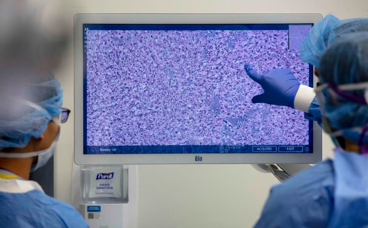
Imagine a surveillance camera that can spot troublemaking cells in your brain before they cause serious problems. This is now a reality with the help of artificial intelligence (AI) and advanced microscopy. Scientists have developed a technique that can detect cancer cells, like those from glioblastomas, deep in brain tissue.
Researchers from EMBL and Heidelberg University have introduced AI to a cutting-edge microscope, allowing them to see and track specific cells with unmatched clarity. This breakthrough helps scientists study glioblastoma cells, one of the most dangerous brain cancers, and could lead to better ways to diagnose the disease early.
A New Microscopy Method
In 2021, EMBL researchers and collaborators from around the world developed a new microscopy technique. This approach helps scientists see neurons and other brain cells deep within regions like the cortex and hippocampus, where it was previously difficult to observe due to scattered light.
The new method, which adjusts for light distortion, has wide-ranging applications in brain research. The microscope can now track living neurons and other cells in the brain for long periods, thanks to custom AI enhancements. These AI tools help distinguish different parts of the cells’ environment, offering crucial insights into cell behavior.
Testing the Technique
Varun Venkataramani, a researcher at Heidelberg University Hospital, saw potential in this technique for studying glioblastomas, which often invade white matter areas like the corpus callosum. This method allowed his team to observe tumor cells in these critical brain regions for the first time, providing valuable insights into how the tumors spread.
AI Enhances Imaging
The AI component significantly improved the microscope’s performance by reducing noise in the images and distinguishing between different structures, such as myelinated fibers and blood vessels. This enhancement is key for identifying tumor cells within their complex environment.
The research team hopes to continue refining this technology to make it suitable for clinical use, potentially improving how brain tumors are diagnosed and treated in the future. This development shows promise in bridging the gap between laboratory research and clinical practice, thanks to strong interdisciplinary collaboration.
Source: technologynetworks